Understanding the Frozen Section Procedure: A Closer Look at Intraoperative Pathology
The Frozen Section Procedure is a diagnostic test used predominantly in surgical pathology to provide a rapid microscopic specimen analysis during surgery. This technique plays a crucial role in guiding
The Frozen Section Procedure is a diagnostic test used predominantly in surgical pathology to provide a rapid microscopic specimen analysis during surgery. This technique plays a crucial role in guiding surgeons to make immediate decisions about the extent of surgery needed based on the presence or absence of disease. The procedure is named "frozen" because the tissue sample is quickly frozen, which allows thin sections to be cut and examined under a microscope.
Why it’s done-
The primary reason for performing a frozen section analysis is to provide immediate diagnostic information that can influence the course of surgery. Here are some of the critical situations in which it is employed:
Cancer Surgery: While removing a tumor, surgeons must ensure they have excised all cancerous tissue while preserving as much healthy tissue as possible. Frozen section analysis helps determine if the edges (margins) of the removed tissue are free of cancer cells, indicating that the tumor has been completely excised.
Determination of Tissue Type: In some cases, the nature of the lesion or mass is unclear before surgery. A frozen section can help determine whether the tissue is benign or malignant, aiding the surgeon in deciding the extent of surgery needed.
Sentinel Lymph Node Biopsy: In certain cancers, such as breast cancer, a sentinel lymph node biopsy is performed to check if the cancer has spread to the lymph nodes. A frozen section of the sentinel lymph node can provide quick information about the presence of cancer cells.
Surgery Procedure -
The frozen section begins when a surgeon identifies a tissue of interest during surgery and requires immediate pathological examination. The tissue sample is quickly sent to the pathology lab, undergoing a rapid freezing process. This is achieved using compounds like liquid nitrogen or a cryostat machine, freeing the tissue solid enough to be sliced into fragile sections.
These thin sections are then mounted on slides, stained with dyes to highlight different cell structures, and examined under a microscope by a pathologist. The entire process, from the receipt of the specimen to the delivery of results, can be accomplished in approximately 20 to 30 minutes, providing vital information without significant interruption to the ongoing surgery.
Applications -
The primary application of the frozen section procedure is in oncologic surgery, where it helps in determining the nature of a tumor - benign or malignant - and the margins of the tumor. If the margins are clear of cancer cells, the surgeon may conclude the procedure, knowing the tumor has been entirely excised. If cancer cells are found at the edges of the specimen, additional tissue may be removed during the same surgical session.
Apart from oncology, frozen section analysis is used in various other surgical contexts, such as organ transplant surgery, to assess the viability of the donor organ or in endocrine surgery to examine parathyroid glands or thyroid nodules.
Risk and Complications -
While the Frozen Section Procedure is invaluable, it is not without risks and complications. These include:
False Negatives/Positives: The possibility of misdiagnosis, although rare, can lead to inadequate removal of diseased tissue or unnecessary removal of healthy tissue.
Sampling Error: The small sample size may not represent the overall nature of the disease, leading to incorrect conclusions.
Technical Limitations: The quality of frozen sections may not match that of permanently processed tissues, potentially affecting the diagnosis accuracy.
Advantages and Limitations
The frozen section procedure offers the significant advantage of immediate results, which can be crucial in surgical decision-making. It allows for real-time adjusting of surgical plans, potentially sparing patients from additional surgeries. However, it has limitations. Rapidly freezing can sometimes distort the tissue architecture, making interpretation challenging. Additionally, not all tissue types are suitable for frozen section analysis, and in some cases, a more thorough examination using traditional methods may be necessary after surgery.
At Medipulse Hospital, Jodhpur, integrating the frozen section procedure into our surgical pathology services underscores our commitment to providing state-of-the-art care. Our dedicated team of pathologists and surgeons collaborates closely, utilizing this rapid diagnostic tool to ensure precise and timely surgical interventions. The availability of frozen section analysis at Medipulse Hospital enhances our ability to make intraoperative decisions that are informed, patient-centric, and tailored to achieve the best possible outcomes. This reflects our overarching mission to blend innovative medical practices with compassionate care, ensuring every patient receives the highest standard of treatment. For more information about Frozen section, visit the hospital’s website or can contact on 8239345635.
चिरंजीवी योजना में अस्थि रोग के अंतर्गत कूल्हों का प्रत्यारोपण (THR-Total hip replacement) का इलाज़ : महत्वपूर्ण जानकारी और आवेदन प्रक्रिया
कूल्हों का प्रत्यारोपण एक प्रकार की सर्जरी है जिसमें कूल्हे के जोड़ को कृत्रिम भागों से बदल दिया जाता है। यह उपचार तब किया जाता है जब कूल्हे का जोड़ घिस जाता है या अत्यधिक दर्द और अक्षमता का कारण बनता है।
कूल्हों का प्रत्यारोपण एक प्रकार की सर्जरी है जिसमें कूल्हे के जोड़ को कृत्रिम भागों से बदल दिया जाता है। यह उपचार तब किया जाता है जब कूल्हे का जोड़ घिस जाता है या अत्यधिक दर्द और अक्षमता का कारण बनता है। THR उन रोगियों के लिए एक आशा की किरण है जिन्हें गठिया, अस्थि भंग या अन्य कूल्हे की समस्याओं के कारण चलने-फिरने में कठिनाई होती है। कई ऐसे लोग है जो इलाज़ की कमी के कारन चल नहीं पाते और बहुत कम उम्र में उनका चलना - फिरना बंद हो जाता है।
चिरंजीवी योजना एक ऐसी योजना है जिसमे सभी तरह के लोग अपना कोई भी इलाज़ फ्री में करा सकते है। कूल्हों के प्रत्यारोपण का इलाज़ भी इस योजना में फ्री में हो सकता है।
चिरंजीवी योजना में कूल्हों का प्रत्यारोपण (THR) के लिए आवश्यक दस्तावेज:
योजना में लाभ लेने के लिए मरीज का जनाधार कार्ड चालू होना अनिवार्य है। पात्रता मानदंड और आवेदन प्रक्रिया के बारे में संबंधित CMHO विभाग से संपर्क करे ।
अगर मरीज किसी अनुमोदित अस्पताल में चिरंजीवी योजना से कूल्हों का प्रत्यारोपण करवाना चाहते है तो उन्हें उसी दिन अपने जनाधार कार्ड और आधार कार्ड होने की सूचना चिरंजीवी काउंटर पर स्वास्थ्य मार्गदर्शक को सूचित करना अनिवार्य होगा।
जिस बिमारी / डॉक्टर से चिरंजीवी योजना में इलाज़ होना है वो अस्पताल में चिरंजीवी योजना से जुड़ा होना जरूरी है
डॉक्टर की पर्ची होना अनिवार्य है जिसमे-
डॉक्टर द्वारा मरीज के बीमारी का डायग्नोसिस,
साथ ही उसकी कंप्लेंट हिस्ट्री और पूर्व में कोई बीमारी है तो उसके बारे में स्पष्ट रूप से लिखा होना चाहिए,
मरीज को होने वाली सर्जरी के साथ-साथ इम्प्लांट का विवरण होना चाहिए ,
चिरंजीवी के कोड एवं डॉक्टर के सील (RMC No.) और हस्ताक्षर होना भी अनिवार्य है
कूल्हों का प्रत्यारोपण सर्जरी के लिए डॉक्टर द्वारा लिखित मरीज के बीमारी के अनुसार जाँचो (जैसे कूल्हे का X-Ray या MRI) का होना ज़रूरी है। बिना जाँचो के चिरंजीवी में इलाज लेना संभव नहीं है।
जन आधार, आधार और मेडिकल डाक्यूमेंट्स में मरीज का नाम एक ही होने चाहिए। नाम अगर मेल नहीं होता तो मरीज को एक एफिडेविट जमा करवाना अनिवार्य होगा।
कूल्हे के प्रत्यारोपण के लिए अस्पताल का Fully NABH Accrediation होना जरूरी होता है।
आवेदन प्रक्रिया:
1. सबसे पहले मरीज के अस्पताल द्वारा बिमारी से सम्बंधित दस्तावेज देखे जाते है। जिसमे पूर्ण डॉक्टर की पर्ची एवं बीमारी से संबंधित जाँचो पर डॉक्टर के सील और हस्ताक्षर साथ ही डॉक्टर का रजिस्ट्रेशन नंबर (RMC) लिखा होना अनिवार्य है।
2. चिरंजीवी के अंतर्गत अलग अलग बीमारियों के इलाज दिए जाते है। मरीज को बीमारी के अनुसार भर्ती से पूर्व जांचे करवाना अनिवार्य है। इको एवं अन्य रेडियोलोजी रिपोर्ट्स के साथ उनकी फिल्म होना भी अनिवार्य है।
बीमारियों की सूचि के साथ उसमे होने वाली जाँचो की सूचि के लिए आप चिरंजीवी की ऑफिसियल साइट पर देख सकते है।
3. मरीज के भर्ती के समय चिरंजीवी योजना के अंतर्गत, मरीज की फोटो एवं बायोमेट्रिक की प्रक्रिया की जाती है। इसमें अस्पताल द्वारा मरीज के दस्तावेज पोर्टल के माध्यम से आगे चिरंजीवी में भेजे जाते है। पोर्टल पर मरीज़ की सामान्य स्थति होने पर आगे से प्री अप्रूवल मिलने के बाद ही मरीज को उपचार दिया जाता है (इमरजेंसी के केस में अप्रूवल की प्रतीक्षा नहीं की जाती है।)। अप्रूवल मिलने का समय लगभग 2 से 4 घंटे रहता है और अधिकतम 12 घंटे रहता है। भर्ती की यह पूरी प्रक्रिया एक मरीज के लिए लगभग 15 से 20 मिनट रहती है।
4. डिस्चार्ज के समय अस्पताल द्वारा दस्तावेज तैयार किये जाते है जिन्हे मरीज की लाइव फोटो एवं डॉक्टर के साथ भी मरीज की फोटो पोर्टल पर अपलोड की जाती है। साथ ही मरीज का फीडबैक फॉर्म मरीज द्वारा भरवाकर उसे पोर्टल पर अपलोड किया जाता है।
क्या करें
डॉक्टर परामर्श जरूर करवाए । डॉक्टर परामर्श होने के बाद ही मरीज को इलाज़ के लिए योजना के अंतर्गत भर्ती करना संभव हो पायेगा।
योजना के अंतर्गत केवल भर्ती रहने पर ही इलाज मुफ्त होता है ।
इलाज के लिए मरीज को जाँचे भर्ती से पूर्व करवाना अनिवार्य है। बिना जाँचो के प्री अप्रूवल लेना संभव नहीं है। जाँच में X-RAY, CT या MRI है तो इनकी रिपोर्ट और फिल्म होना ज़रूरी है, साथ ही संबंधित डॉक्टर के हस्ताक्षर एवं सील के साथ डॉक्टर का रजिस्ट्रेशन नंबर होना भी अनिवार्य है।
मरीज का जनाधार कार्ड और आधार कार्ड होना अनिवार्य है एवं जनाधार कार्ड में आधार कार्ड नंबर के साथ-साथ आधार कार्ड में बायोमेट्रिक व मोबाइल नंबर भी अपडेट होना अनिवार्य है।
योजना का लाभ केवल MMCSBY अनुमोदित अस्पताल में ही लिया जा सकता है ।
क्या न करें
OPD में चिरंजीवी के अंतर्गत कैशलेस सुविधा नहीं दी जाती है।
अपरिपूर्ण जाँचो एवं डॉक्टर की पर्ची के साथ चिरंजीवी योजना के तहत इलाज संभव नहीं है।
चिरंजीवी योजना के तहत मरीज का इलाज निशुल्क होता है। इसमें मरीज को किसी भी प्रकार की राशि जमा करवाने की आवश्यकता नहीं होती है।
जनाधार, आधार और मेडिकल डाक्यूमेंट्स में मरीज का नाम एक ही होने चाहिए, अलग-अलग दर्ज़ न करवाए।
इस योजना के अंतर्गत इलाज लेने के लिए मरीज को धैर्य रखना आवश्यक है क्योकि इसमें अस्पताल को दस्तावेजों की प्रक्रिया में समय लग सकता है। किसी भी प्रकार की हिंसा अस्पताल कर्मचारियों के साथ न करे।
मेडिपल्स अस्पताल का आर्थोपेडिक विभाग अस्थि रोगों के निदान, उपचार, और निवारण में विशेषज्ञता रखता है। इस विभाग में मरीजों को व्यापक देखभाल और सहायता प्रदान की जाती है, जिसमें चिकित्सा विशेषज्ञ और स्वास्थ्य सेवा प्रदाता शामिल हैं। मेडिपल्स अस्पताल ने चिरंजीवी योजना के तहत राज्य के कई नागरिकों को मुफ्त में कूल्हे के प्रत्यारोपण की सुविधा प्रदान की है। अस्थि रोगों के इलाज में इस अस्पताल की विशेषज्ञता है, और इस योजना के अंतर्गत कई मरीजों का इलाज निःशुल्क किया गया है। चिरंजीवी योजना के अंतर्गत मुफ्त कूल्हे के प्रत्यारोपण के लिए मेडिपल्स अस्पताल में आप इस नंबर 8239345635 पर संपर्क कर सकते हैं।
चिरंजीवी योजना में अस्थि रोग के अंतर्गत घुटनो का प्रत्यारोपण (TKR-Total knee replacement) का इलाज़ : महत्वपूर्ण जानकारी और आवेदन प्रक्रिया
घुटने का दर्द, आज के समय में, विशेषकर बुजुर्ग जनसंख्या में एक सामान्य समस्या बन गई है। इसका मुख्य कारण है अस्थि रोग जिसके चलते घुटनों का कार्टिलेज धीरे-धीरे घिसता चला जाता है, और अंततः, घुटनों का प्रत्यारोपण (Total Knee Replacement - TKR) एकमात्र विकल्प बचता है।
घुटने का दर्द, आज के समय में, विशेषकर बुजुर्ग जनसंख्या में एक सामान्य समस्या बन गई है। इसका मुख्य कारण है अस्थि रोग जिसके चलते घुटनों का कार्टिलेज धीरे-धीरे घिसता चला जाता है, और अंततः, घुटनों का प्रत्यारोपण (Total Knee Replacement - TKR) एकमात्र विकल्प बचता है। घुटनों का प्रत्यारोपण एक सर्जिकल प्रक्रिया है जिसमें घुटनों के क्षतिग्रस्त हिस्सों को निकालकर उनकी जगह कृत्रिम भागों से बदला जाता है। यह उपचार उन रोगियों के लिए अत्यंत लाभदायक सिद्ध होता है, जिनके घुटनों में गंभीर दर्द और चलने-फिरने में कठिनाई होती है। कई ऐसे लोग है जो पैसो की कमी की वजह से तकलीफ पाते रहते है और अपने घुटनो का इलाज़ नहीं करा पाते है। चिरंजीवी योजना, एक सरकारी योजना है, जिसका उद्देश्य है आर्थिक रूप से कमजोर वर्गों को उच्च स्तरीय और महंगे मेडिकल ट्रीटमेंट तक पहुँच प्रदान करना। इस योजना के तहत, घुटनों का प्रत्यारोपण जैसे जटिल और महंगे उपचार फ्री में संभव है।
चिरंजीवी योजना में घुटनो का प्रत्यारोपण (TKR) के लिए आवश्यक दस्तावेज:
योजना में लाभ लेने के लिए मरीज का जनाधार कार्ड चालू होना अनिवार्य है। पात्रता मानदंड और आवेदन प्रक्रिया के बारे में संबंधित CMHO विभाग से संपर्क करे ।
अगर मरीज किसी अनुमोदित अस्पताल में चिरंजीवी योजना से घुटनो के प्रत्यारोपण करवाना चाहते है तो उन्हें उसी दिन अपने जनाधार कार्ड और आधार कार्ड होने की सूचना चिरंजीवी काउंटर पर स्वास्थ्य मार्गदर्शक को सूचित करना अनिवार्य होगा।
जिस बिमारी / डॉक्टर से चिरंजीवी योजना में इलाज़ होना है वो अस्पताल में चिरंजीवी योजना से जुड़ा होना जरूरी है
डॉक्टर की पर्ची होना अनिवार्य है जिसमे-
o डॉक्टर द्वारा मरीज के बीमारी का डायग्नोसिस,
o साथ ही उसकी कंप्लेंट हिस्ट्री और पूर्व में कोई बीमारी है तो उसके बारे में स्पष्ट रूप से लिखा होना चाहिए,
o मरीज को होने वाली सर्जरी के साथ-साथ इम्प्लांट का विवरण होना चाहिए ,
o चिरंजीवी के कोड एवं डॉक्टर के सील (RMC No.) और हस्ताक्षर होना भी अनिवार्य है
o घुटनो का प्रत्यारोपण सर्जरी के लिए डॉक्टर द्वारा लिखित मरीज के बीमारी के अनुसार जाँचो (जैसे घुटने का X-Ray या MRI) का होना ज़रूरी है। बिना जाँचो के चिरंजीवी में इलाज लेना संभव नहीं है।
जन आधार, आधार और मेडिकल डाक्यूमेंट्स में मरीज का नाम एक ही होने चाहिए। नाम अगर मेल नहीं होता तो मरीज को एक एफिडेविट जमा करवाना अनिवार्य होगा।
घुटने के प्रत्यारोपण के लिए अस्पताल का Fully NABH Accrediation होना जरूरी होता है, साथ-साथ मरीज़ का 1 महीने पहले से ही इलाज़ चलता हुआ होना चाहिए और 2 अस्थि रोग विशेषज्ञ (एक सरकारी अस्पताल और दूसरा निजी अस्पताल) से घुटनो के प्रत्यारोपण के लिए परामर्श किया हुआ होना अनिवार्य है।
आवेदन प्रक्रिया:
सबसे पहले मरीज के अस्पताल द्वारा बिमारी से सम्बंधित दस्तावेज देखे जाते है। जिसमे पूर्ण डॉक्टर की पर्ची एवं बीमारी से संबंधित जाँचो पर डॉक्टर के सील और हस्ताक्षर साथ ही डॉक्टर का रजिस्ट्रेशन नंबर (RMC) लिखा होना अनिवार्य है।
चिरंजीवी के अंतर्गत अलग अलग बीमारियों के इलाज दिए जाते है। मरीज को बीमारी के अनुसार भर्ती से पूर्व जांचे करवाना अनिवार्य है। इको एवं अन्य रेडियोलोजी रिपोर्ट्स के साथ उनकी फिल्म होना भी अनिवार्य है।
बीमारियों की सूचि के साथ उसमे होने वाली जाँचो की सूचि के लिए आप चिरंजीवी की ऑफिसियल साइट पर देख सकते है।
मरीज के भर्ती के समय चिरंजीवी योजना के अंतर्गत, मरीज की फोटो एवं बायोमेट्रिक की प्रक्रिया की जाती है। इसमें अस्पताल द्वारा मरीज के दस्तावेज पोर्टल के माध्यम से आगे चिरंजीवी में भेजे जाते है। पोर्टल पर मरीज़ की सामान्य स्थति होने पर आगे से प्री अप्रूवल मिलने के बाद ही मरीज को उपचार दिया जाता है (इमरजेंसी के केस में अप्रूवल की प्रतीक्षा नहीं की जाती है।)। अप्रूवल मिलने का समय लगभग 2 से 4 घंटे रहता है और अधिकतम 12 घंटे रहता है। भर्ती की यह पूरी प्रक्रिया एक मरीज के लिए लगभग 15 से 20 मिनट रहती है।
डिस्चार्ज के समय अस्पताल द्वारा दस्तावेज तैयार किये जाते है जिन्हे मरीज की लाइव फोटो एवं डॉक्टर के साथ भी मरीज की फोटो पोर्टल पर अपलोड की जाती है। साथ ही मरीज का फीडबैक फॉर्म मरीज द्वारा भरवाकर उसे पोर्टल पर अपलोड किया जाता है।
क्या करें
डॉक्टर परामर्श जरूर करवाए । डॉक्टर परामर्श होने के बाद ही मरीज को इलाज़ के लिए योजना के अंतर्गत भर्ती करना संभव हो पायेगा।
योजना के अंतर्गत केवल भर्ती रहने पर ही इलाज मुफ्त होता है ।
इलाज के लिए मरीज को जाँचे भर्ती से पूर्व करवाना अनिवार्य है। बिना जाँचो के प्री अप्रूवल लेना संभव नहीं है। जाँच में X-RAY, CT या MRI है तो इनकी रिपोर्ट और फिल्म होना ज़रूरी है, साथ ही संबंधित डॉक्टर के हस्ताक्षर एवं सील के साथ डॉक्टर का रजिस्ट्रेशन नंबर होना भी अनिवार्य है।
मरीज का जनाधार कार्ड और आधार कार्ड होना अनिवार्य है एवं जनाधार कार्ड में आधार कार्ड नंबर के साथ-साथ आधार कार्ड में बायोमेट्रिक व मोबाइल नंबर भी अपडेट होना अनिवार्य है।
योजना का लाभ केवल MMCSBY अनुमोदित अस्पताल में ही लिया जा सकता है ।
क्या न करें
OPD में चिरंजीवी के अंतर्गत कैशलेस सुविधा नहीं दी जाती है।
अपरिपूर्ण जाँचो एवं डॉक्टर की पर्ची के साथ चिरंजीवी योजना के तहत इलाज संभव नहीं है।
चिरंजीवी योजना के तहत मरीज का इलाज निशुल्क होता है। इसमें मरीज को किसी भी प्रकार की राशि जमा करवाने की आवश्यकता नहीं होती है।
जनाधार, आधार और मेडिकल डाक्यूमेंट्स में मरीज का नाम एक ही होने चाहिए, अलग-अलग दर्ज़ न करवाए।
इस योजना के अंतर्गत इलाज लेने के लिए मरीज को धैर्य रखना आवश्यक है क्योकि इसमें अस्पताल को दस्तावेजों की प्रक्रिया में समय लग सकता है। किसी भी प्रकार की हिंसा अस्पताल कर्मचारियों के साथ न करे।
मेडिपल्स अस्पताल का आर्थोपेडिक विभाग अस्थि रोगों के निदान, उपचार, और निवारण में विशेषज्ञता रखता है। इस विभाग में मरीजों को व्यापक देखभाल और सहायता प्रदान की जाती है, जिसमें चिकित्सा विशेषज्ञ और स्वास्थ्य सेवा प्रदाता शामिल हैं। मेडिपल्स अस्पताल ने चिरंजीवी योजना के तहत राज्य के कई नागरिकों को मुफ्त में घुटने के प्रत्यारोपण की सुविधा प्रदान की है। अस्थि रोगों के इलाज में इस अस्पताल की विशेषज्ञता है, और इस योजना के अंतर्गत कई मरीजों का इलाज निःशुल्क किया गया है। चिरंजीवी योजना के अंतर्गत मुफ्त घुटने के प्रत्यारोपण के लिए मेडिपल्स अस्पताल में आप इस नंबर 8239345635 पर संपर्क कर सकते हैं।
PET CT scan side effects : Understanding the risks and complications.
PET CT scan is a diagnostic imaging test that combines positron emission tomography (PET) and computed tomography (CT) to produce highly detailed images of the body.
PET CT (Positron Emission Tomography - Computed Tomography) is a medical imaging technique that combines the use of radioactive tracers with CT scans to produce detailed images of internal body structures. While PET CT scans are generally considered safe, like any medical procedure, they do come with some risks and potential side effects. In this article, we will discuss some of the possible PET CT scan side effects.
1. Allergic reactions: Some patients may experience an allergic reaction to the radioactive tracer used in PET CT scans. Symptoms may include itching, hives, and difficulty breathing. In rare cases, a severe allergic reaction known as anaphylaxis may occur, which can be life-threatening.
2. Radiation exposure: PET CT scans involve the use of radiation, which can increase the risk of cancer and other health problems over time. However, the amount of radiation used in PET CT scans is generally considered safe.
3. Nausea: Nausea is a common side effect of PET-CT scans, particularly when patients receive contrast material. Some patients may experience nausea or vomiting after a PET CT scan. This is usually mild and goes away on its own within a few hours.
4. Headache: Headaches are a common side effect of PET CT scans. They are usually mild and go away on their own within a few hours.
5. Dizziness: Some patients may experience dizziness or lightheadedness after a PET CT scan. This is usually mild and goes away on its own within a few hours.
6. Pain or discomfort: Some patients may experience pain or discomfort at the injection site or in other parts of the body during or after the PET CT scan.
Conclusion :
while PET CT scans are generally considered safe, they do come with some risks and potential side effects. It is important to discuss these risks with your doctor before undergoing a PET CT scan.
At Medipulse Hospital, our experienced medical professionals take all necessary precautions to minimize these risks and ensure that our patients receive the best possible care. If you have any concerns or questions about PET CT scan or any other medical procedure, our team is always available to provide you with the information and support you need.
Understanding PET CT Scan - How it works and what to expect
PET CT (Positron Emission Tomography - Computed Tomography) is a medical imaging technique that combines the functional information of PET and the anatomical details of CT to produce highly detailed images of the human body.
PET CT (Positron Emission Tomography - Computed Tomography) is a medical imaging technique that combines the functional information of PET and the anatomical details of CT to produce highly detailed images of the human body. During the scan, a small amount of radioactive tracer is injected into the patient's bloodstream, which is then absorbed by the organs and tissues being imaged. As the tracer decays, it emits positrons which collide with electrons and produce gamma rays. These gamma rays are detected by the PET scanner and used to create a three-dimensional image of the body's metabolic activity. The CT scanner then produces a detailed anatomical image of the same area.
Techniques used in PET CT Scan:
The following are the techniques used in PET-CT scan:
Radiotracer injection: Before the scan, a small amount of radioactive material known as a radiotracer is injected into the patient. The radiotracer is usually a sugar molecule tagged with a radioactive atom.
PET imaging: The radiotracer emits positrons, which collide with electrons in the body, producing gamma rays that are detected by a PET scanner. PET imaging produces three-dimensional images of the distribution of the radiotracer in the body, indicating areas of high metabolic activity.
CT imaging: A CT scan is then performed immediately after the PET scan. The CT scan uses X-rays to produce detailed images of the internal structures of the body. This provides anatomic information that helps to identify the location of abnormal metabolic activity detected by the PET scan.
Fusion imaging: The PET and CT images are combined using special software to create fused images. The fused images provide a more accurate and comprehensive view of the body, enabling physicians to identify and locate abnormalities.
Image interpretation: The fused images are interpreted by a radiologist or a nuclear medicine specialist, who looks for areas of abnormal metabolic activity that may indicate the presence of cancer or other diseases.
Conclusion:
Medipulse Hospital is equipped with state-of-the-art PET CT scanning technology, and our highly trained staff ensures that every patient is comfortable and well-informed before, during, and after the scan. Our hospital follows strict safety protocols to ensure that the scan is performed with the utmost care, and our team of radiologists is always available to interpret the results and provide appropriate treatment recommendations. If you need a PET CT scan, we encourage you to schedule an appointment with us and experience the high-quality care that we provide.
Benefits of ICE therapy
Ice therapy, also known as cryotherapy, is a popular treatment for sports injuries. It involves the application of ice or a cold pack to an injured area of the body to reduce pain, swelling, and inflammation. Ice therapy
Ice therapy, also known as cryotherapy, is a popular treatment for sports injuries. It involves the application of ice or a cold pack to an injured area of the body to reduce pain, swelling, and inflammation. Ice therapy is an inexpensive, safe, and effective method for managing sports injuries, and it has been widely used by athletes and trainers for many years. Here are some of the key benefits of ice therapy:
Reduces Inflammation: Inflammation is a normal response to injury, but it can cause pain and swelling. Ice therapy can help reduce the amount of inflammation in an injured area, reducing pain and swelling and promoting healing.
Reduces Pain: Cold temperatures have a numbing effect on the skin, reducing the sensation of pain. Ice therapy can help to relieve pain from sports injuries and make it easier to move the affected area.
Improves Healing: Ice therapy can help to improve circulation and reduce inflammation, which can speed up the healing process. Cold temperatures also help to slow down the metabolic rate, reducing the demand for oxygen and nutrients, allowing the body to focus on repairing the damaged tissue.
Prevents Tissue Damage: Ice therapy can help to prevent tissue damage by reducing the amount of blood flow to the affected area. This can help to reduce the risk of further injury and promote healing.
Affordable and Convenient: Ice therapy is a low-cost and convenient method of treating sports injuries. Ice packs are readily available and can be easily stored in a freezer, making it easy to access when needed.
In conclusion, ice therapy is a simple and effective way to manage sports injuries. It can help to reduce pain, swelling, and inflammation, improve healing, and prevent tissue damage. If you have a sports injury, it is recommended to consult a doctor or physical therapist to determine the best treatment plan for your injury. Ice therapy may be a part of your rehabilitation program, along with other treatments such as physical therapy, medications, and bracing.
What is knee arthroscopy and what are the precautions you should keep
Knee arthroscopy is a common surgical procedure used to diagnose and treat various knee conditions such as torn cartilage, osteoarthritis, and ligament damage. As with any surgery, proper postoperative
Knee arthroscopy is a common surgical procedure used to diagnose and treat various knee conditions such as torn cartilage, osteoarthritis, and ligament damage. As with any surgery, proper postoperative care is essential to ensure a successful recovery and prevent any complications. If you or a loved one has recently undergone knee arthroscopy, it is important to follow the following precautions to promote a safe and quick healing process.
Rest and Ice: After the surgery, it is crucial to rest and elevate your affected knee as much as possible. You can also apply ice packs to help reduce swelling and pain. However, be sure to avoid applying ice directly to the skin, as it can cause discomfort and damage.
Physical Therapy: Your doctor will most likely prescribe physical therapy to help you regain your range of motion and strengthen your knee. It is important to attend all physical therapy sessions as directed and follow the exercises and instructions provided by your therapist.
Medications: Your doctor may prescribe pain medications to help manage your postoperative pain. Take these medications as directed, and do not exceed the recommended dosages. If you experience any side effects, be sure to inform your doctor immediately.
Weight Management: It is important to avoid putting any unnecessary stress on your knee during the recovery process. Try to avoid carrying heavy objects and maintain a healthy weight to minimize the strain on your knee.
Avoid High-Impact Activities: High-impact activities, such as running and jumping, can put unnecessary strain on your knee and slow down the healing process. Avoid these activities and stick to low-impact exercises such as swimming or cycling until you have been cleared by your doctor.
Follow Your Doctor's Instructions: Your doctor will provide you with specific postoperative instructions, including information on when you can return to work, drive, and resume physical activity. It is important to follow these instructions carefully to ensure a smooth recovery.
In conclusion, taking proper precautions after knee arthroscopy is crucial for a successful recovery and to minimize the risk of any complications. If you experience any pain, swelling, or any other symptoms that concern you, be sure to contact your doctor immediately. With the right care and attention, you should be able to return to your normal activities in no time.
5 Ways You Can Reduce/Control Your Cholesterol Levels
High cholesterol levels lead to cardiac problems and clogged arteries. It is one of the most common health concerns nowadays, with 6 out of 10 people having abnormal cholesterol levels. High cholesterol levels, or,
High cholesterol levels lead to cardiac problems and clogged arteries. It is one of the most common health concerns nowadays, with 6 out of 10 people having abnormal cholesterol levels. High cholesterol levels, or, to be more precise high LDL levels (bad cholesterol), are one of the leading causes of cardiac diseases to become the number one cause of death among human beings worldwide. So, the risk is real, so if you have high cholesterol levels, follow these five ways to reduce or control your cholesterol levels.
5 Ways To Control/Reduce Your Cholesterol Levels
Active Lifestyle
To start reducing your cholesterol levels, you need to begin with getting an active lifestyle. The key here is to go out, live your life, enjoy physical activity and make the most of your days. A sedentary lifestyle with little physical activity is a root cause of many health problems, including high cholesterol levels. So, if you want to reduce or control your LDL levels, start a more active lifestyle; how do you do that? The answer’s in the next point.
Exercise Regularly
Be it going for a walk or hitting the gym. Exercising is crucial to stay healthy. Lifting weights is optional, but cardio is not. You must work at least a few hours a week to ensure your health is at peak levels. This will help you maintain your overall physical health and keep your cholesterol levels in check. Also, exercising can help you maintain your BMI, which is crucial. Why? Let’s discuss the next point.
Maintaining A Healthy BMI
To ensure that your body is in the best shape possible, tracking and maintaining your BMI can greatly help. It ensures that your body weight is appropriate per your age and you are as healthy as possible. Additionally, keeping your weight in check is a great way to ensure your body is healthy and free from other health problems. And if you are following the previous steps already, you don’t have to do anything else to maintain weight.
Eat Healthy Consistently
Don’t eat junk food, reduce consumption of red meat and fried food, eat some vegetables, try having a diverse range of food and eat a balanced diet overall. You need to take care of these things to keep your cholesterol levels in control. And additionally, you have to eat like this consistently to ensure you get the nutrients needed to be in top form physically.
No Smoking
Smoking can do no good for you. It’s ideal if you don’t already; if you do, it’s in your best interest to quit. You can take the help of several medications to deal with the withdrawal symptoms, but you should contemplate quitting first. Smoking adversely affects your lungs, heart, and overall health. So, quitting smoking even without doing the above can still help you be healthier.
Conclusion
These are five ways you can control or reduce your cholesterol levels. Obviously, there are more ways, and you can check out Medipulse hospital to learn more about cholesterol. They are experts in treating and helping people with cardiac problems. MediPulse is a multispeciality hospital with experienced doctors and staff that can help you live a better life and recover from cardiac illnesses. For more information about cholesterol treatment at MediPulse website, you can check out the hospital’s website or visit Medipulse hospital in Jodhpur.
Cholesterol: The Ultimate Cardiac Health Regulator
Cholesterol is not a bad thing. It helps your body produce healthy cells. However, given their waxy nature, if they are above the recommended levels, they can cause cardiac health problems. The main problem with
Cholesterol is not a bad thing. It helps your body produce healthy cells. However, given their waxy nature, if they are above the recommended levels, they can cause cardiac health problems. The main problem with Cholesterol is that you must go through regular health checkups and blood work to ensure optimum levels.
You also need to regulate your diet and maintain an active lifestyle to maintain a healthy cholesterol level as you age. These factors make it one of the leading causes of cardiac problems in people of advanced age. So, let’s dive deep into why Cholesterol is dangerous, how to test abnormal cholesterol levels, and how to treat it.
Problem With High Cholesterol
High cholesterol levels cause cholesterol deposits inside the blood vessels and constrict blood flow. This causes clogged arteries, which result in a reduction in the blood flow to the heart. As a result, patients can become susceptible to heart attacks, strokes, and other cardiovascular diseases, all of which can be fatal. So, if not monitored regularly and kept in check, high cholesterol levels can cause serious heart health issues that can threaten your lifespan. Now that you know the problems, let’s learn how to help you monitor abnormal cholesterol levels.
Monitoring Abnormal Cholesterol Levels
Even high and abnormal levels of Cholesterol don’t come with any bodily symptoms. Blood work is the only way to determine your cholesterol levels. These tests should be conducted regularly to ensure your levels are in check. For people who are at risk of cardiac diseases due to genetics or lifestyle should get blood work done more often to keep a check on cholesterol levels.
According to doctors, cholesterol levels should be checked from a young age starting around ten, and done once every five years. As you age, the frequency needs to be increased as much as once every year. If discrepancies are found in your reports, doctors may advise you to get your blood work done every few months. But why do high cholesterol levels exist in the first place? Let’s focus on the causes of abnormal cholesterol levels.
Causes Of Abnormal Cholesterol Levels
Cholesterol is divided into two parts; there is LDL and HDL. LDL is low-density lipoprotein, and HDL is high-density lipoprotein. LDL is bad Cholesterol, and if you have an abnormal level of LDL, you have high Cholesterol. So, the idea behind Cholesterol is that when in excess, LDL transports the cholesterol particles to different parts of your body and deposits them there.
Abnormal levels of LDL can be caused by a sedentary lifestyle combined with excessive consumption of alcohol and smoking. Additionally, genetic factors can also come into play, which may make you genetically predisposed to getting an abnormal cholesterol level. Let’s focus now on how to treat abnormal cholesterol levels.
How To Treat Abnormal Cholesterol Levels?
Cholesterol levels themselves don’t cause any harm to your body, and they are key in causing cardiac diseases, which can harm your health. So, to treat abnormal cholesterol levels, you need to maintain a healthy heart. Here are some ways you can maintain your heart health.
Regular exercising
Keeping your BMI in check
A steady and healthy diet
Abstaining from smoking and drinking alcohol
Try and manage your stress levels
Conclusion
Cholesterol is a silent killer and can seriously affect your health if not monitored correctly. So, if you are an adult, make sure you get your blood work done at least once a year to ensure your cholesterol levels are normal. You should also follow the steps mentioned above to maintain a healthy heart. And if you have high cholesterol levels, feel free to visit MediPulse hospital in Jodhpur for the best cardiac care.
What Are Chronic Ulcers & How Do You Treat Them?
Chronic ulcers/wounds are open wounds on the surface of your skin that take a long time to heal. These ulcers are common on your ankles, feet, and legs. There are several reasons that can cause these ulcers.
Chronic ulcers/wounds are open wounds on the surface of your skin that take a long time to heal. These ulcers are common on your ankles, feet, and legs. There are several reasons that can cause these ulcers. Given that the treatment is a long process, here’s all you need to know about chronic ulcers and their treatment process in Jodhpur.
What Are Chronic Ulcers/Wounds?
Chronic ulcers are wounds on your legs that, due to tissue loss, look like a hole inside your body. As they are open, they can become infected and get worse if not treated accordingly. Even with treatment, these wounds take a long time to heal. These wounds are not a result of accidents; they are caused by varying factors, which are discussed below.
What Causes Chronic Ulcers?
There are several risk factors that can lead to chronic ulcers. Let’s review the risk factors and why they contribute to chronic ulcers.
Chronic Venous Insufficiency
Due to blockages in the veins or due to reflux, which is the backflow of blood inside the veins, tissues in your leg can be affected by a condition known as chronic venous insufficiency. It is caused due to prolonged exposure to high pressure inside the veins of your legs. This is one of the main causes of chronic ulcers and wounds.
Obesity
Obesity is another leading cause of chronic ulcers because obesity has adverse effects on your circulatory system. The added pressure on your veins and arteries, combined with the possibility of developing clots in them, are what make obesity a leading cause of chronic ulcers.
Diabetes Mellitus
Diabetes is also a common cause of chronic ulcers because of its inherent effect on your body. Diabetes makes your body prone to infections, especially in your foot. Any wound takes longer to heal if you have diabetes. So, the possibility of getting chronic wounds increases with having diabetes mellitus.
It should be noted that chronic ulcers may seem like a variation of skin cancer, but for the most part, ulcers on the leg are not skin cancer. They are chronic ulcers or wounds.
How Do You Treat Chronic Ulcers/Wounds?
The treatment of chronic ulcers depends greatly on finding out the root cause of it. These ulcers don’t have any go-to treatment option particularly. The goal is to find the main cause and treat that until the ulcer is healed. So, for you to get treatment for your chronic ulcers/wounds, you need to see a specialist, a vascular surgeon, to be precise.
A vascular surgeon will thoroughly examine your body to determine the cause of your ulcer. It’s crucial that you disclose your entire medical history to them for a correct diagnosis. If you go through any of the conditions listed above that are causes of ulcers, you should let your doctor know. Other than that, if you have any other vascular disease, rheumatoid arthritis, or varicose veins, you should let your doctor know.
With the right information, your doctors can figure out the root cause and treat that, which will ultimately help your chronic ulcer heal.
Conclusion
That’s a brief overview of chronic ulcers and how to treat them. If you or someone you know has chronic ulcers/wounds, feel free to visit MediPulse hospital in Jodhpur. MediPulse is the best tertiary care multispeciality hospital in Jodhpur, with ample experience in dealing with chronic ulcer cases. For more information about chronic ulcer treatment at MediPulse, visit the hospital website or visit the hospital for consultation.
What Is Gynecomastia? Do You Need Treatment For It? What Are Your Treatment Options?
As the quality of life improves in India, people take time to try and fix the quality of life problems for a better body image. Body dysmorphia is a real issue, and even though women more commonly go through it,
As the quality of life improves in India, people take time to try and fix the quality of life problems for a better body image. Body dysmorphia is a real issue, and even though women more commonly go through it, it affects men as well. Gynecomastia is a condition that doesn’t hurt your physical health, but it can definitely trigger body dysmorphia in your head.
Given this condition is faced by men, it often goes unnoticed and untreated even after triggering body dysmorphia in men. So, if you or someone in your known circle is facing Gynecomastia in Jodhpur, this blog will help you get the right treatment approach if needed.
What Is Gynecomastia?
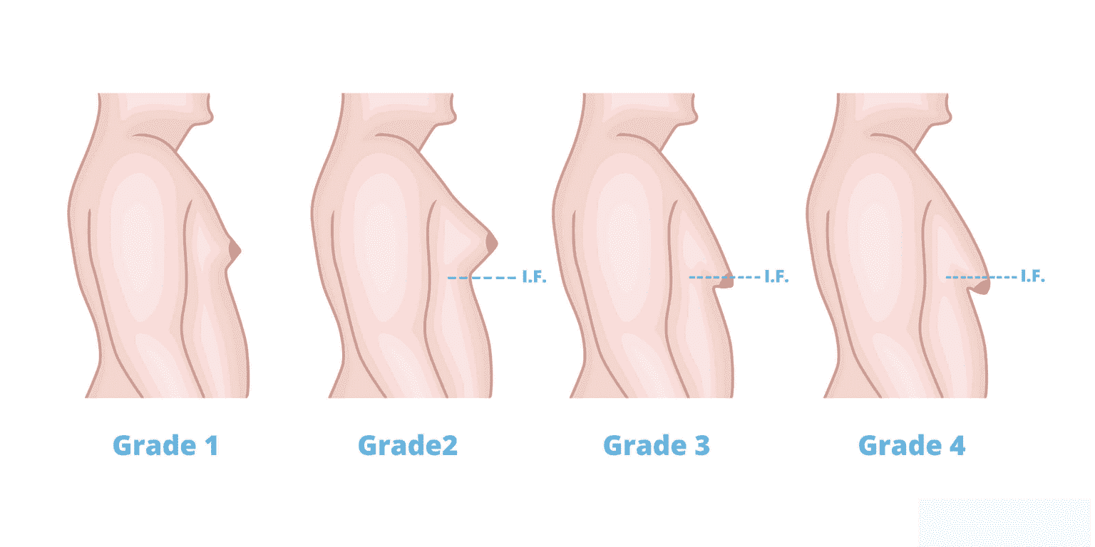
Gynecomastia is a condition that men suffer from where the breast gland tissues start growing abnormally due to hormonal imbalances. This condition can be triggered at any point in a man’s life due to imbalances in their estrogen and testosterone levels. It can affect the two breasts differently and cause abnormal growth in the tissues of one or both breasts.
There is another condition known as pseudogynecomastia, which refers to a fat increase in the breast area of men or boys. This issue is not caused by hormonal imbalance but rather a matter of gaining weight. So, now that you know the types of gynecomastia, let’s discuss its causes and if you need treatment for it.
Causes Of Gynecomastia
Gynecomastia is caused when estrogen surpasses the testosterone level in a man’s body. The condition can be caused by several factors, such as diabetes, poor diet, overusing steroids, or chronic stress. Generally, gynecomastia doesn’t come with any symptoms, but in some cases, the changes in size can cause pain or tenderness in the breasts or discharge to come out of the nipples. In such cases, you should visit a doctor. Otherwise, treatment is not necessary unless it creates a problem for your body image or self-confidence.
Treatment Options For Gynecomastia
If you are suffering from pain or discharge from your breasts, doctors will, in most cases, try to treat the root cause of your problem. These treatments can take time, and you can’t guarantee that your breast size will be reduced.
Treatment options include testosterone supplements to help you boost your testosterone levels. You can also be asked to review your diet or diabetic status in order for a thorough examination of the cause of your issue. If you are found to have diabetes, treatment plans will be utilized accordingly to help reduce your diabetic levels and keep them under control; the same goes for a poor diet.
The last resort treatment is to go for gynecomastia surgery or male breast reduction surgery. The doctors will remove the additional breast tissues in your body through surgery to help you get a reduced breast size and a better look. As this is a non-essential surgery except in specific cases, most medical insurance will not cover the cost of the surgery and the recovery.
There are very few risks to the surgery, and it doesn’t even require you to have an overnight hospital stay unless explicitly requested. Based on your condition, doctors can suggest that you go through with the surgery, or you can even request the doctors for the surgery yourself.
Conclusion
This is a brief overview of gynecomastia. It is most common nowadays among adult and aging men in the age group between 50 to 80. So, if you are going through this issue, you can visit MediPulse hospital for a checkup to find out the root cause behind your problem and help treat it accordingly. The experienced staff and doctors at MediPulse hospital have considerable experience dealing with gynecomastia surgery if required. So, for further information and treatment, visit MediPulse hospital, Jodhpur.
Scarring: What Is It?, Its Types, & Treatments
Scars, the things that remind you that you got hurt, or the things that supposedly make you more manly. Whatever your take on scars might be, medically, they are tissues formed at the sight of any cuts or bruises
Scars, the things that remind you that you got hurt, or the things that supposedly make you more manly. Whatever your take on scars might be, medically, they are tissues formed at the sight of any cuts or bruises on your body. Any break or cut on your skin can lead to Scarring, but it doesn’t always. If you develop scars or not, that can be affected by the site of the wound, its depth, and its size. So, now that you know the factors that make a scar and look for its types and treatment options in Jodhpur, this blog will help you further.
Are There Different Types Of Scars?
Yes, there are four types of scars. The name of the scar also kind of defines the way you get them. It’s important to note before moving forward that scars are inherently untreatable. You can do things to reduce the appearance of scars, but it’s quite difficult to fully treat a scar. There are several things that play into it, which will be discussed in the next section dedicated to treatments. For now, let’s explore the four types of scars.
Acne Scars
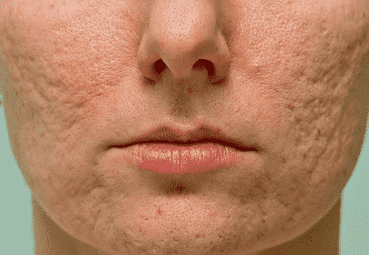
Acne is common among young adults and teenagers, but all types of acne don’t lead to scars. Typically, only severe acne cases lead to Scarring. The appearance of the scars can also differ based on different issues, but severe cases often lead to deep and pit-like scars. In some cases, they can even be angular or wavelike in appearance. Treating these scars depends on the type and cause of your acne.
Contracture Scars
You can get these scars from serious burns. The scar tissues tighten your skin and restrict your ability to move that part of your body. These scars also go deep and can even go as deep as your muscles and nerves. Contracture scars are serious and need intensive medical care. As you would imagine, these scars can’t heal themselves, and treatment options are also quite limited for them, given they are very serious injuries.
Hypertrophic Scars
These scars are formed at the site of an injury. They are also easy to identify because they are red, and they don’t expand beyond the site of the wound; this is important because that’s how you can differentiate it from a Keloid scar, which will be explained next. Hypertrophic scars are treatable with steroids and silicone sheets. However, some appearance of the scar might still remain after the treatments as well.
Keloid Scars
Keloid scars can occur over a number of injuries; they are caused by your body’s healing process, which, when it becomes aggressive, creates keloid scars. These scars can go beyond the boundaries of the wound, and like contracture scars, they can prohibit your ability to move. These scars can be stopped from forming using gel packs and silicone sheets after an injury. Once you have them, treatment is less helpful but can still work towards reducing the appearance of the scar.
Treatment Options
There are mainly three types of treatments when it comes to scars. They are topical, surgical, and injections. Let’s dive into each of them.
Topical treatment is anything that has to be applied externally on your scar. It is effective in cases where the scars are not very serious or if they are from anything like plastic surgery. Topical treatment includes ointments, lotions, and even oral medications for dealing with the itchiness of your scars.
Surgical treatment is for serious scars where topical treatment doesn’t work effectively. Surgery aims to remove the scar’s appearance through techniques such as laser treatment or dermabrasion. In serious cases, skin grafting is also commonly used to treat scars, especially ones that limit your ability to move.
Lastly, steroid injections are also a common way of treating Scarring, especially keloid and hypertrophic scars.
Conclusion
These are all the types of scars and their treatment options. If you want to learn more about scars, you can check out MediPulse hospital’s website or visit them in Jodhpur for a quick consultation.


